What is confocal microscopy?
The confocal microscope scans a laser beam through a sample to excite molecules that are in focus. The molecules then emit photons, which are measured by a detector. A pinhole in front of the detector blocks out-of-focus light and hence improves image quality.
Confocal microscopy offers an improved resolution and signal-to-noise ratio over widefield. The ability to vary the height of the focal plane simultaneously while experimenting is also remarkably useful for 3D reconstruction of larger specimens. This method is more time-consuming than widefield in acquisition and image production and generally higher maintenance, in some cases there is also a tradeoff where some resolution is sacrificed to distinguish different structures in a given sample. Confocal Microscopy is an excellent method of optical imaging often preferred by researchers for applications involving visualization of genetic material or structural components of cells. It enables the assessment of the colocalization of structures and has had large ramifications in studies of the viability of cells.

Three color confocal image of Cas9 binding to a single DNA molecule tethered between two optically trapped beads. Blue/green/red markers indicate the position of individual Cas9 complexes.
Explore other imaging techniques
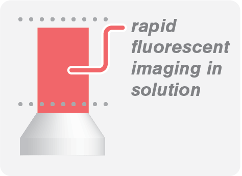
Widefield
Image biomolecular processes with low protein concentration in solution with high acquisition rates.
STED Super Resolution
The perfect choice for performing experiments in highly crowded environments, offering unprecedented resolution (< 35 nm).
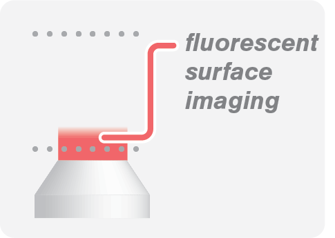
TIRF Fluorescence
Suitable for visualization on surfaces as it eliminates background fluorescence outside the focal plane.
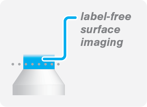
IRM (label-free)
IRM allows you to visualize microtubules without the need for fluorescence labeling.
Our solution
The C-Trap® Optical Tweezers – Fluorescence & Label-free Microscopy is the world’s first instrument that allows simultaneous manipulation and visualization of single-molecule interactions in real time. It combines high resolution optical tweezers, fluorescence and label-free microscopy and an advanced microfluidics system in a truly integrated and correlated solution.
The C-Trap offers you a fast workflow to seamlessly catch and manipulate single molecules. The instrument measures their structural changes or interactions while you visualize them in teal time with high spatial and temporal resolution, ultimately offering you a complete and detailed picture of biomolecular properties and interactions.
Curious to learn more?
