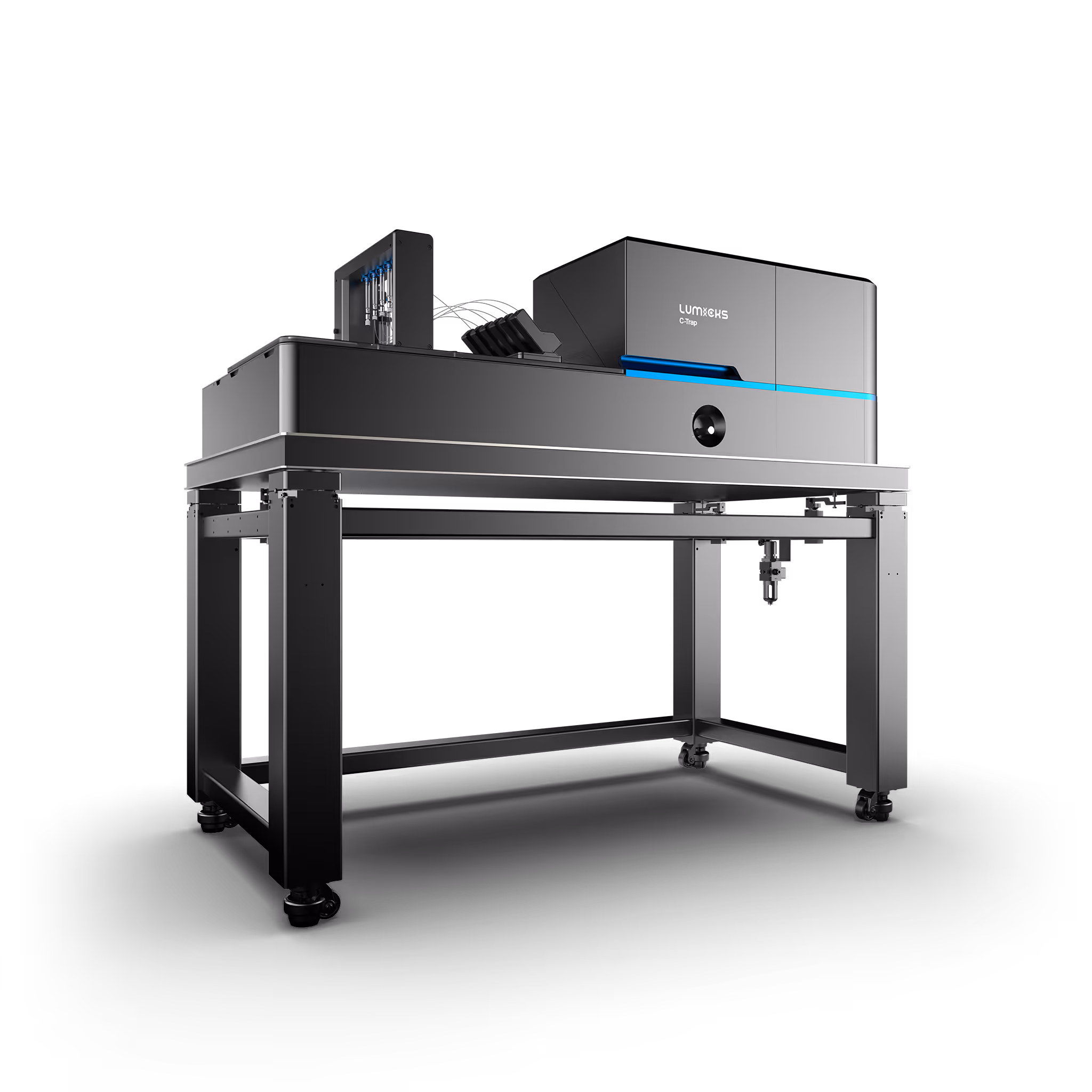
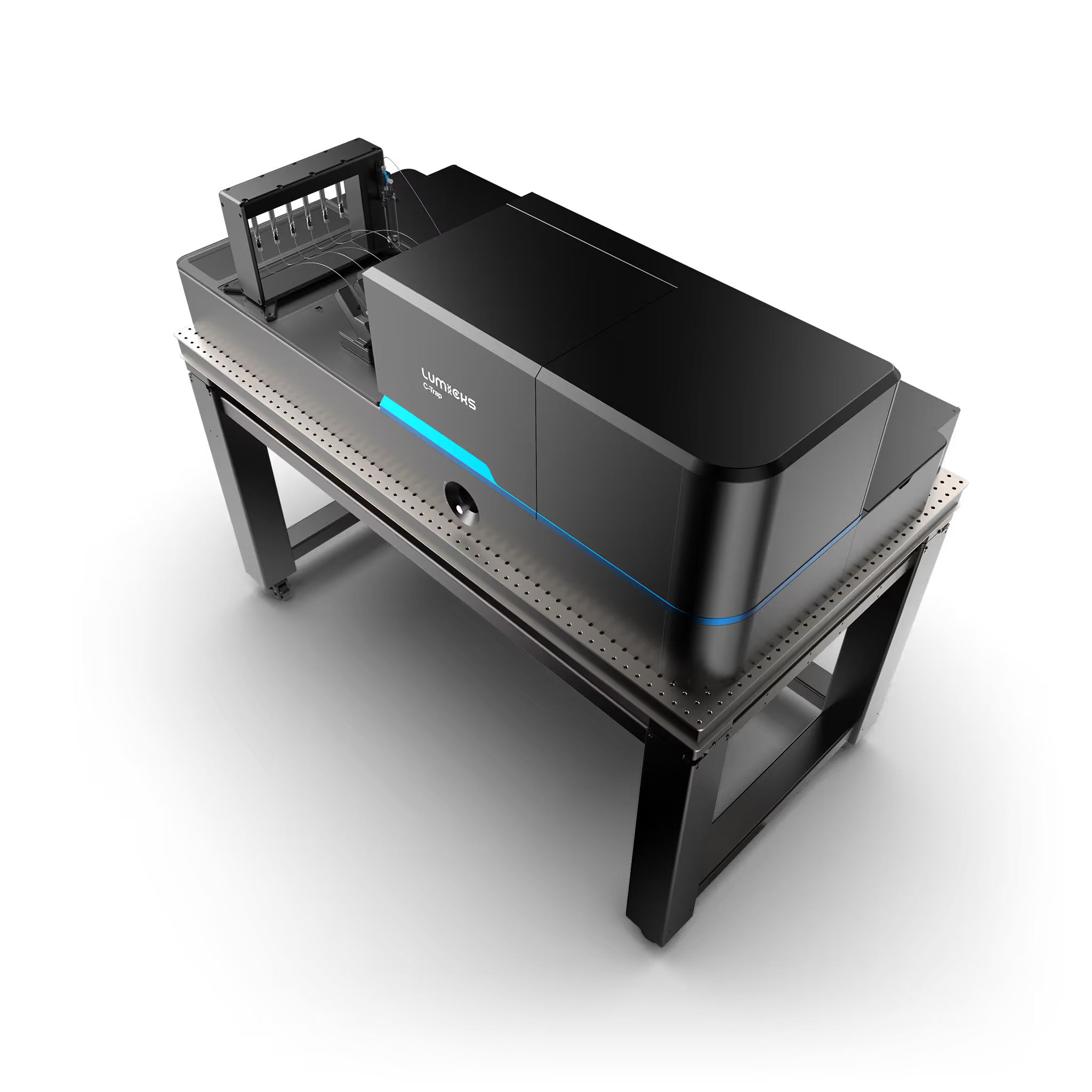
C-Trap
Molecular biology as never seen before
The world’s first dynamic single-molecule microscope to allow simultaneous manipulation and visualization of single-molecule interactions in real time.
Highly detailed results at high throughput
Manipulate your sample with exceptional stability and precision, utilizing powerful automation features and extensions.
Easy to learn workflow & software
Start experimenting right away using our conventional microfluidics and software solutions designed specifically for C-Trap experiments.
Purpose-fit for your needs
Choose from a wide degree of optical trap and imaging wavelength configurations to fit your workflow.
Workflow video
A typical experiment
Watch our short workflow video to understand what a typical day with a C-Trap looks like

0:00
Workflow
High-throughput experiment workflow
From sample loading to data analysis in less than 30 minutes.
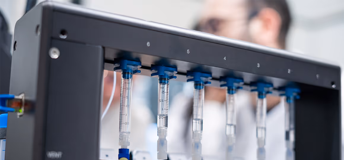
Load your samples
Load your samples and conditions into the syringes of the 5-channel automated microfluidics system. Pipetting each reagent takes seconds thanks to the twist-and-lock syringe adaptor, with which you can quickly and easily refill individual syringes.

Assemble your assay without physical barriers
Seamlessly move the optical traps within the laminar flowcell to catch beads, move them between the 5 microfluidics lanes, and assemble your constructs.

Perform and automate your experiments
Gather statistically relevant quantities of data and publishable results with ease. The C-Trap allows for automation with full access to all relevant system parameters and data streams.
With an experimental run taking less than 80 seconds, including automated assay assembly, it's possible to get 18 useable sets of data within half an hour.

Organize and analyze your data
View, compare, and export your fully correlated data streams. The C-Trap stores all your metadata so you never lose valuable information, and always have the option to reproduce your experiments.
Our analysis software comes with tutorials and sample notebooks that can serve as a scaffold for your own analyses. In addition, we have an open-source platform where you can upload, download, and review user scripts for free.
Platform
Take a look inside
Discover the manipulation and imaging technologies that bring the C-Trap to life
Precise sample manipulation
Study the smallest of interactions using optical traps combined with advanced software features. Available in multiple configurations, the C-Trap can handle a broad variety of assays.
Multicolor fluorescence imaging
Visualize biological processes such as protein kinetics and (un)binding events on DNA or measure conformational changes of proteins by combining the C-Trap with FRET. Our fast 1D scanning capabilities make it excellent for constructs such as DNA or filaments.
Laminar microfluidics
Assemble your assay and study a variety of proteins or conditions in the same experiment with our our dedicated laminar flow cell and automated microfluidics solution.
Customer testimonials
Trusted by visionary scientists in molecular biology

Priya's team reveals how proteins shape diseases like neurodegeneration and cancer
"The application of C-Trap, which offers a truly correlative single-molecule force spectroscopy and fluorescence microscopy, will enable us to interrogate how single-molecule properties of individual IDP chains are correlated with their phase behavior."
Priya Banerjee, PhD
Professor of Biophysics
University of Buffalo

Stephen studies how cells repair broken DNA to prevent cancer and genetic diseases
"The combination of structural data, EM data and single-molecule studies provided three powerful tools to elucidate the functions of the BCDX2 complex (a key DNA repair regulator) in DNA repair and cancer avoidance."
Stephen West, PhD
Senior Group Leader
The Francis Crick Institute

Shannon's group explores how physical forces shape cell division and cell-cell communication
"LUMICKS really helped me by making a turn-key instrument that is just good enough that [has] all the fancy capability I need, and I do not need to worry about optimizing alignment or changing things."
Shannon Yan, PhD
Assistant Professor
Stanford University
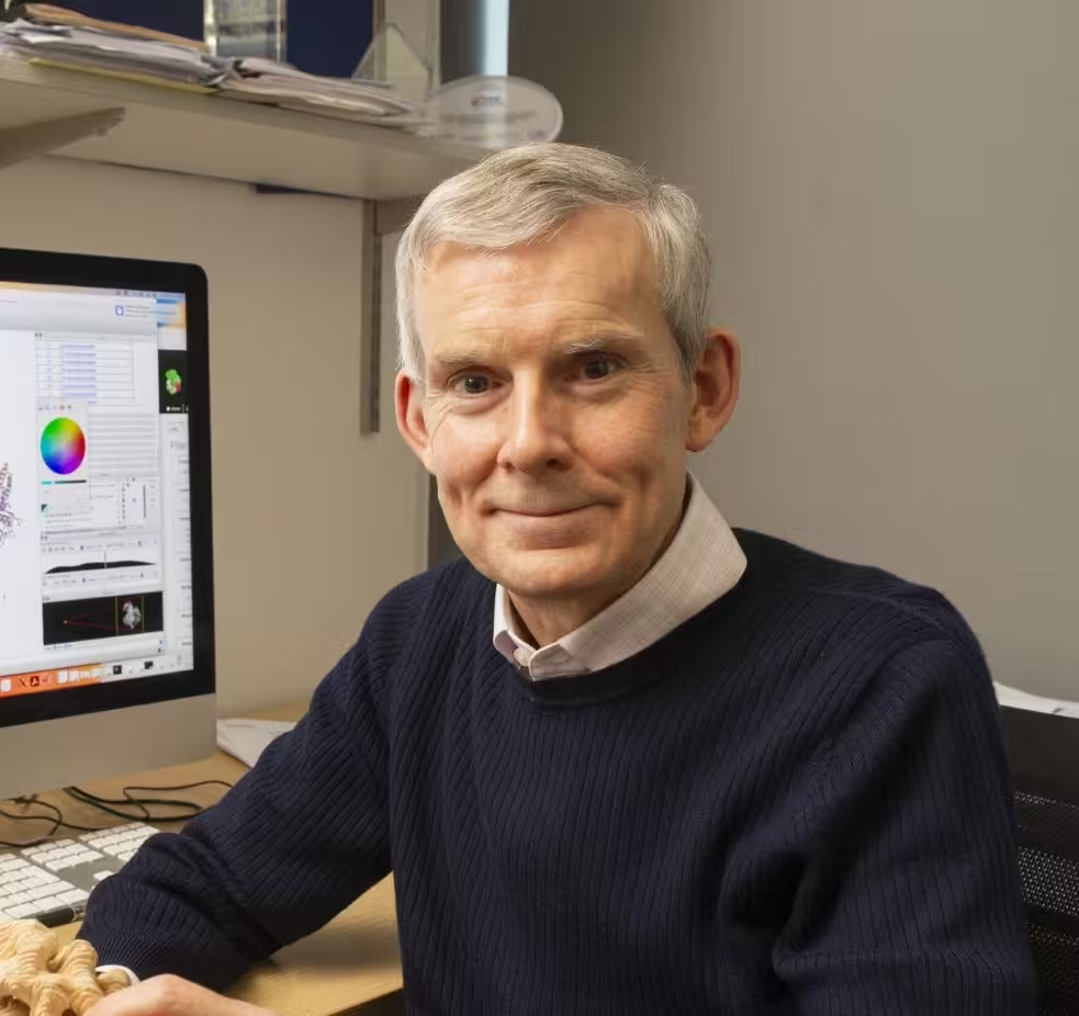
David's group works on understanding how cells prevent cancer and genetic errors
"We are dedicated to understanding important biological processes at the molecular level to tackle major problems in human health and disease. We welcome LUMICKS’ C-Trap technology into our laboratory and we look forward to utilizing it in a wide range of research programs."
David Barford, PhD
Group Leader
MRC Laboratory of Molecular Biology
Software
Data acquisition and analysis tools for every type of (single) molecule
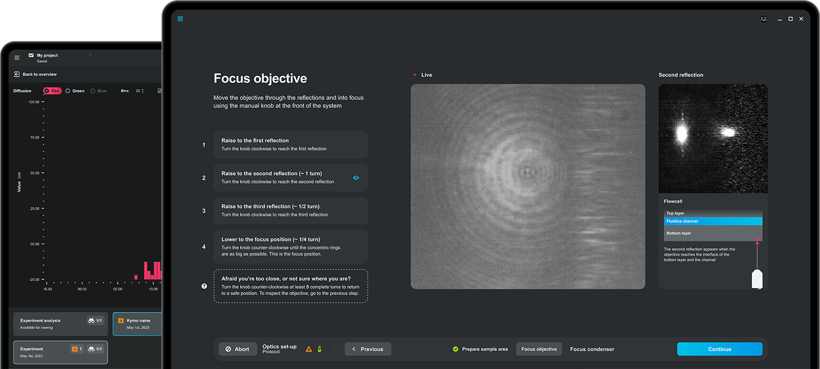
Execute and analyze your C-Trap experiments using our intuitive Bluelake & Lakeview software.
Execute and automate your experiment with ease
Whether you are navigating through the laminar flow cell, controlling the optical traps, or utilizing our powerful automation features, our easy to use Bluelake solution puts you in control.
Adapt and iterate on the fly with real-time data
Bluelake presents you with a full overview of your experiment’s data as it gets measured. Catch new insights quickly and adapt your experiment on the fly to make the most out of your time.
Obtain immediate insights and study them wherever, whenever
Acquire statistical insights across multiple Dynamic Single-Molecule measurements in only a few clicks. Lakeview makes it easy to gain reliable information quickly and integrate into your own pipeline.
Curious about our data analysis solution? Take a look at our dedicated Lakeview page.
Key content
Download the brochure
Get the latest all-in-one overview
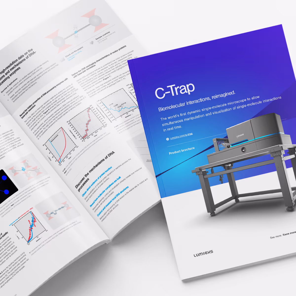
C-Trap Product Brochure
Brochure
The latest all-in-one overview of C-Trap's features, options, and specifications.
Now available
C-Trap Accelerator Suite
Supercharge your C-Trap into a faster, easier, more reproducible powerhouse. C-Trap Accelerator Suite boosts the capabilities of the instrument you already own, letting you capture publishable single-molecule data in half the time with rock-solid reproducibility.
Benefits
Beyond the product
Take a look at the perks that come with being one of our users

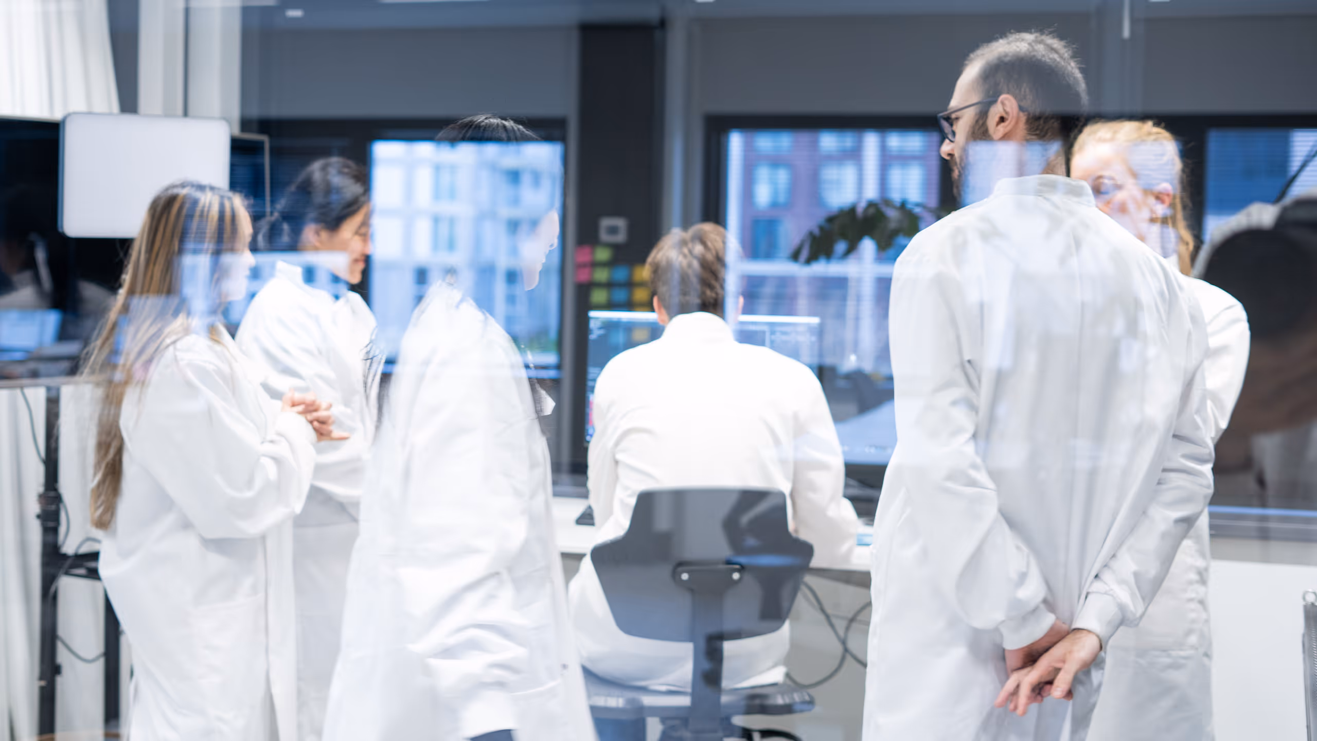
Symposia
Join the community
SMBIO (Single-molecule Biology) is LUMICKS’ symposia format. We regularly host events across the world, aiming to bring together scientists interested in dynamic single-molecule applications to share their research, connect, and exchange ideas and experiences.

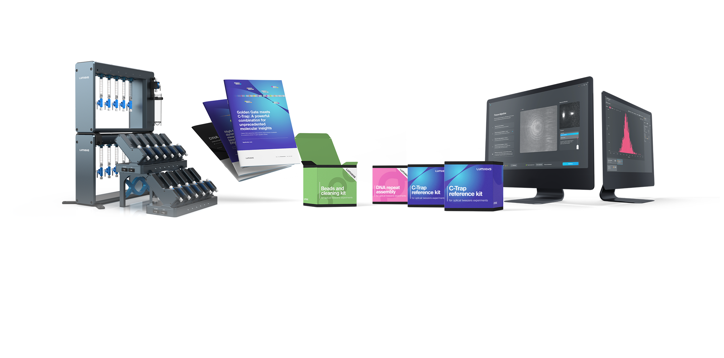
Store
Ready-made reagent kits and tailored sample preparation services
Use our biochemistry platform and internal expertise for the purification, labeling, and preparation of your reagents for optical trapping experiments. With our multidisciplinary expertise in molecular biology, biochemistry, and dynamic single-molecule analysis, we’ve created the most extensive selection of ready-made reagents, kits, and tailored services to prepare your samples.
Support & service
Your success is our success
Our goal is to minimize your time to results, ensure success, and provide instruments that are easy to use and maintain. To this end we have dedicated teams providing user training, application support and system check-ups.
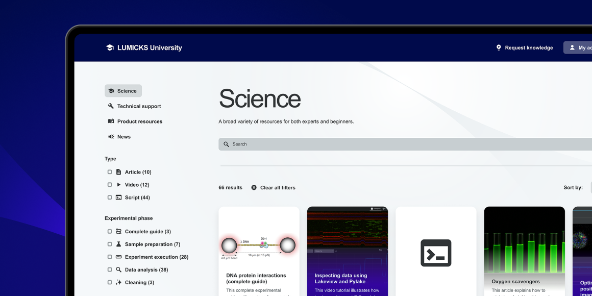
LUMICKS University
Your gateway to mastering single-molecule experiments
LUMICKS University is our all-in-one learning hub to simplify your learning, deepen your knowledge and accelerate your discoveries, designed exclusively for LUMICKS customers.
Adopting Dynamic Single-Molecule
Let’s begin your LUMICKS journey
Our experts are ready to learn about your research challenges and see where our technologies can bring value.
Demonstrating value
Our application scientists can help create interest among potential users through organizing different events such as seminars, workshops, and demonstrations, as well as meet with stakeholders individually to understand and help solve their needs.
Grant & tender support
Throughout our history we have supported multiple successful grants across a broad spectrum of funding and users involved. Our application scientists are experienced in highlighting the unique value of Dynamic Single-molecule and its solutions, and are able to collect proof-of-concept data to strengthen your grant application.