Dynamic Single-Molecule
Revealing biomolecular insights never before available
What if you could measure molecular properties and interactions while simultaneously revealing the processes in real-time? A technology that unravels the crucial and dynamic interactions taking place at the molecular level and gives you direct proof of the mechanisms involved. Our Dynamic Single-Molecule technology provides you with all of that, enabling the understanding of the root of disease development at the molecular level and accelerate therapeutic breakthroughs.
Gain unprecedented insights into the complex mechanistic details of interactions
Correlate structure and function like no other technology can
Get started in no time through accessible workflows and solutions
The challenge
Molecular mechanisms are at the heart of understanding biology
All biological processes ultimately take place at the molecular level, all diseases arise at this level, and practically all drugs act at this level. However, many life science and tools that are used today to approximate and extract molecular function such as structural biology, bulk functional assays, cell imaging, and localization assays, give either detailed structural or functional information - but rarely both.
Unable to observe molecular processes in real time, they often fail to reveal crucial mechanistic details of the molecular factors at play. This lack of direct observation of the underlying dynamic processes often results in ambiguous data, not suited to deliver clear insights.

A solution for tomorrow
Dynamic Single-Molecule, the missing link
Dynamic Single-Molecule combines live visualization, manipulation, and force measurement at the smallest molecular scale, leads to single-molecule imaging and base-pair resolution measurements of the interactions between biomolecules. Taken together, this provides – for the first time – the crucial dynamic and functional mechanistic information that is complementary to molecular structure, bulk functional assays, and cell imaging.

Workflow
Experiene a typical experiment for yourself
Let’s take you through the various steps involved in an experiment

0:00
Applications
Unravel every interaction between protein and DNA, with unprecedented resolution and speed
Use Dynamic Single-Molecule to study a wide variety of DNA-Binding protein behavior.
DNA Repair
Reveal the dynamics of DNA repair mechanisms
Featuring case studies from:


DNA Organization
Discover the mechanisms and roles of chromatin organization and decipher the epigenetic code
Featuring case studies from:


DNA Replication
Study and visualize DNA replication mechanisms at the nanoscale
Featuring case studies from:


DNA Transcription
Study and visualize DNA transcription mechanisms at the nanoscale
Featuring case studies from:
DNA Editing
Study and visualize DNA editing mechanisms on the nanoscale
Featuring case studies from:


DNA/RNA Structure
Reveal the structural dynamics of RNA & DNA in real time
Featuring case studies from:


Study (un)folding events, phase separation, mechanical properties, and cytoskeletal structures and behavior.
Utilize Dynamic Single-Molecule to study a wide variety of interactions.
Protein Folding
Study protein folding and conformational dynamics at the nanoscale
Featuring case studies from:

Phase Separation
From fundamental mechanism to biological function – explore biomolecular condensates across scales
Featuring case studies from:

Mechanobiology
Uncover mechanical principles of cellular function at the molecular level
Featuring case studies from:
Cytoskeletal Structure and Transport
Study cytoskeletal motors in real-time at the nanoscale
Featuring case studies from:

Solutions
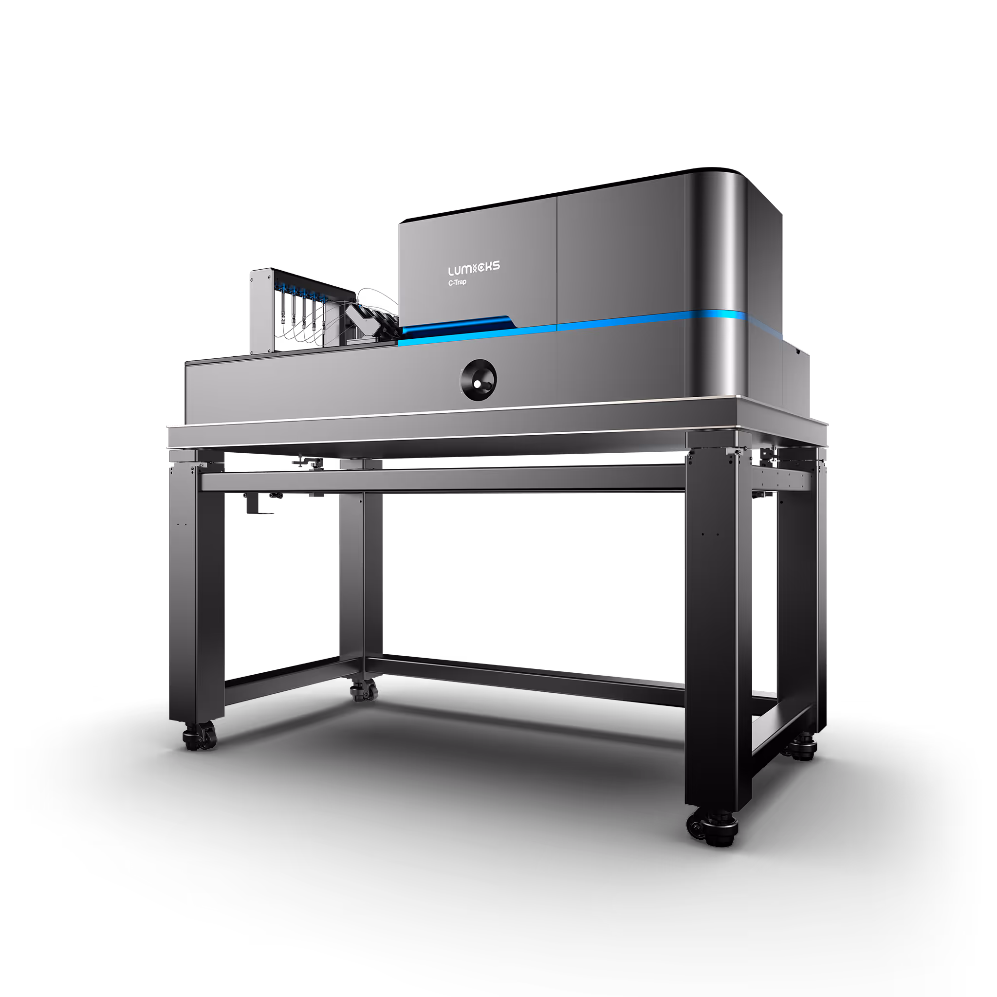
C-Trap
Molecular biology as never seen before
The C-Trap® provides the world’s first dynamic single-molecule microscope to allow simultaneous manipulation and visualization of single-molecule interactions in real time.
Technical note:
Filter DSM shows 3 items
To show 1 or more authors, we are using finsweet attributes v2
The publications detail page has the format for these authors
Filter DSM shows 3 items
To show 1 or more authors, we are using finsweet attributes v2
The publications detail page has the format for these authors
Structural basis of supercoiling-induced CRISPR–Cas9 off-target activity
Smith, Q.M. et al.
2026
Nature
This is some text inside of a div block.
NAC promotes co-translational protein folding at the ribosomal tunnel exit
Santos, J. et al.
2026
Molecular Cell
This is some text inside of a div block.
Tuning the sensitivity of mechanosensory receptors through histidine scanning
Wang, Y. et al.
2026
Cell
This is some text inside of a div block.
Adopting Dynamic Single-Molecule
Let’s begin your LUMICKS journey
Our experts are ready to learn about your research challenges and see where our technologies can bring value.
Demonstrating value
Our application scientists can help create interest among potential users through organizing different events such as seminars, workshops, and demonstrations, as well as meet with stakeholders individually to understand and help solve their needs.
Grant & tender support
Throughout our history we have supported multiple successful grants across a broad spectrum of funding and users involved. Our application scientists are experienced in highlighting the unique value of Dynamic Single-Molecule and its solutions.