A recent study by Szu‐Ning Lin et al. published in Nature Scientific Reports aims at shining light on the dynamics of HU-DNA interactions. The researchers employed the C-Trap®, a dynamic single-molecule method, to visualize in real-time the diffusion and binding of HU molecules along the DNA, while manipulating DNA conformation and changing Mg2+ concentrations. The study for the first time provided direct information on the dynamics of the interaction between HU and DNA and how this interaction is influenced by ionic conditions and DNA conformation.
HU (histone‐like protein from E. coli strain U93) is a well-characterized DNA-binding protein found in most bacteria. It is a member of the architectural DNA–binding protein family that, based on the ability to modify DNA conformation, plays a crucial role in gene expression.
Previous studies reported conflicting results on the mobility of HU on DNA, with a five times higher diffusion coefficient in vitro than in vivo. Possibly, because of different ionic conditions and crowding phenomena. Other studies investigated the effect of salt conditions but didn’t provide direct information on the dynamics of the HU-DNA interaction. This underlines the compelling need for a direct, single-molecule approach to the matter. As the authors say:
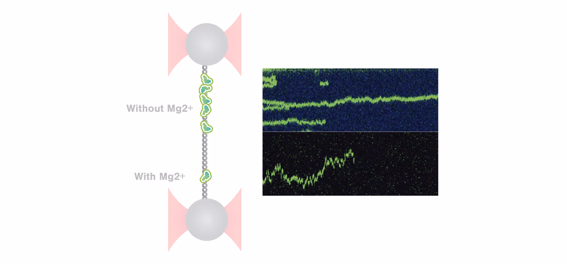
“Using confocal microscopy combined with optical tweezers in a microfluidic system, we visualize and characterize the binding of individual HU dimers on DNA.”
Employing the C-Trap, Szu‐Ning Lin et al. found that in absence of Mg2+ and at applied tension between 10 – 60 pN, HU displays high binding affinity to DNA and low mobility once bound – resulting in HU clustering. Contrary, in presence of Mg2+ and applied tension up to 50 pN, HU shows only low binding to DNA, with high diffusion coefficients and no HU clustering. However, interestingly, applying tension over 50 pN increases the binding affinity. These results suggest that, in presence of Mg2+, the change in DNA conformation (at over 50 pN) leads to a different binding mode.
This work by Szu‐Ning Lin et al. offers for the first time a mechanistic insight into the interaction between HU dimers and the DNA filament with a single-molecule resolution proving the role that ionic strength and DNA conformation play in this process. Observation of these interactions and the different binding mode was not possible with other methods, thus highlighting the value of dynamic single-molecule technology in revealing the details of molecular mechanisms.
For more information on the discovery of Szu‐Ning Lin et al., read the paper published in Nature Scientific Reports with the title: “Direct visualization of the effect of DNA structure and ionic conditions on HU–DNA interactions”.