Documentation
Optical tweezers
Learn all about optical tweezers and what they can do

What are optical tweezers and what can they do?
In 2018 Arthur Ashkin won the Nobel Prize in Physics “for the optical tweezers and their application to biological systems”. In essence, he discovered that light’s momentum could serve as an incredibly sensitive set of tweezers to catch and study biomolecules.
By shooting a laser through a microscope, he created a highly focused light beam, strong enough to trap and “trap” small objects, such as plastic beads. These beads can be coated to stick to various biomolecules, such as DNA, RNA, proteins, or filaments. Two trapped beads can be used as hooks to hold a single molecule on its respective ends. We can then control the lasers and move the beads with the tethered biomolecule and, for instance, stretch it. The ability to manipulate molecules in this way allows us to measure forces (for example, upon stretching) and monitor the beads’ positions. These outcomes can then serve us to calculate the tethered molecule (for example, its elasticity) and study structural transitions.
By shooting a laser through a microscope, he created a highly focused light beam, strong enough to trap and “trap” small objects, such as plastic beads. These beads can be coated to stick to various biomolecules, such as DNA, RNA, proteins, or filaments. Two trapped beads can be used as hooks to hold a single molecule on its respective ends. We can then control the lasers and move the beads with the tethered biomolecule and, for instance, stretch it. The ability to manipulate molecules in this way allows us to measure forces (for example, upon stretching) and monitor the beads’ positions. These outcomes can then serve us to calculate the tethered molecule (for example, its elasticity) and study structural transitions.
Discovery of optical tweezers
First introduced by Arthur Ashkin and colleagues in 1986 at Bell labs, optical tweezers quickly emerged as an indispensable tool that can be used for a variety of different applications in chemistry and biology1.
Not long after this initial breakthrough, optical tweezers, or optical traps as they are otherwise known, were successfully used to physically trap and control viruses, bacteria and single-cells, paving the way for the mechanical and kinetic study of biomolecules at the single-molecule level2-3.
Presently, optical tweezers have been used in a variety of different applications, such as in the study of the interactions between proteins and DNA involved in DNA organization, replication, transcription and repair; the study of the energy landscape of proteins and the kinetics of molecular motors4-7.
Simply put, the value of optical tweezers lies in the fact that they can be used to perform experiments to probe the properties of single-molecules by applying forces in the range of picoNewtons and by measuring distance displacements in the range of nanometers.

Working principle of optical tweezers
Optical tweezers are based on the principle of light carrying momentum proportional to its energy and propagation direction.
When a laser beam passes through an object, it bends and changes direction (called refraction) and alters its momentum. According to Newton’s third law, the object undergoes an equal and opposite momentum change, a reaction force, for the system to conserve the total momentum.
Figure 1 illustrates the transfer of light momentum occurring when a light beam travels through a bead. In a typical optical tweezers configuration, the incoming light originates from a focused laser beam through a microscope objective and focuses on a spot in the sample. The spot subsequently creates a trap able to hold a small object in place.

Figure 1: Re-direction of a light path and change of momentum as it passes through a microsphere or “bead” with high index of refraction related to the medium (left). Momentum of equal and opposite force is transferred from the photons to the bead according to Newton’s Law of energy conservation (right).
Laser trapping of beads
In a typical optical tweezers configuration, the incoming light originates from a focused laser beam through a microscope objective and focuses on a spot in the sample. The spot creates a trap able to hold a small dielectric object at place.
The total forces experienced by the object, or bead in most experimental settings, consist of a scattering force and a gradient force8. The scattering force arises when a light beam is scattered by the surface of the object. This scattering produces a net momentum transfer from the light photons to the object and causes the bead to be pushed towards the beam propagation. The gradient force results from the intensity profile of the laser beam which acts as an attractive force, drawing the bead towards the region with greater light intensity. In the case of a focused laser beam with a Gaussian intensity profile (a normal distribution), the gradient force pulls the object into the center of the focal plane.
The reason the object stays in the center of the beam is because of the sum of the forces acting upon it. In the center, rays of light refract or scatter through the object the same way on both sides of the vertical plane, which cancels forces from moving the object sideways. If the object drifts to one side, it returns to the center. Think of a spring that accelerates back to the center when displaced from its equilibrium position. Figure 2 shows how the gradient force restores an off-centered bead towards the center of the focal plane, eff ectively trapping the object in all dimensions

Figure 2: Two light paths passing through a dielectric micron sized bead. Due to the light gradient, the path originating from the center of the beam carries more photons than the light path commencing from the outlines of the beam, resulting to a larger force pulling the bead towards the focal point.
References
8. Neuman, K. et al.
2004
Review of Scientific Instruments
https://doi.org/10.1063/1.1785844
Solutions
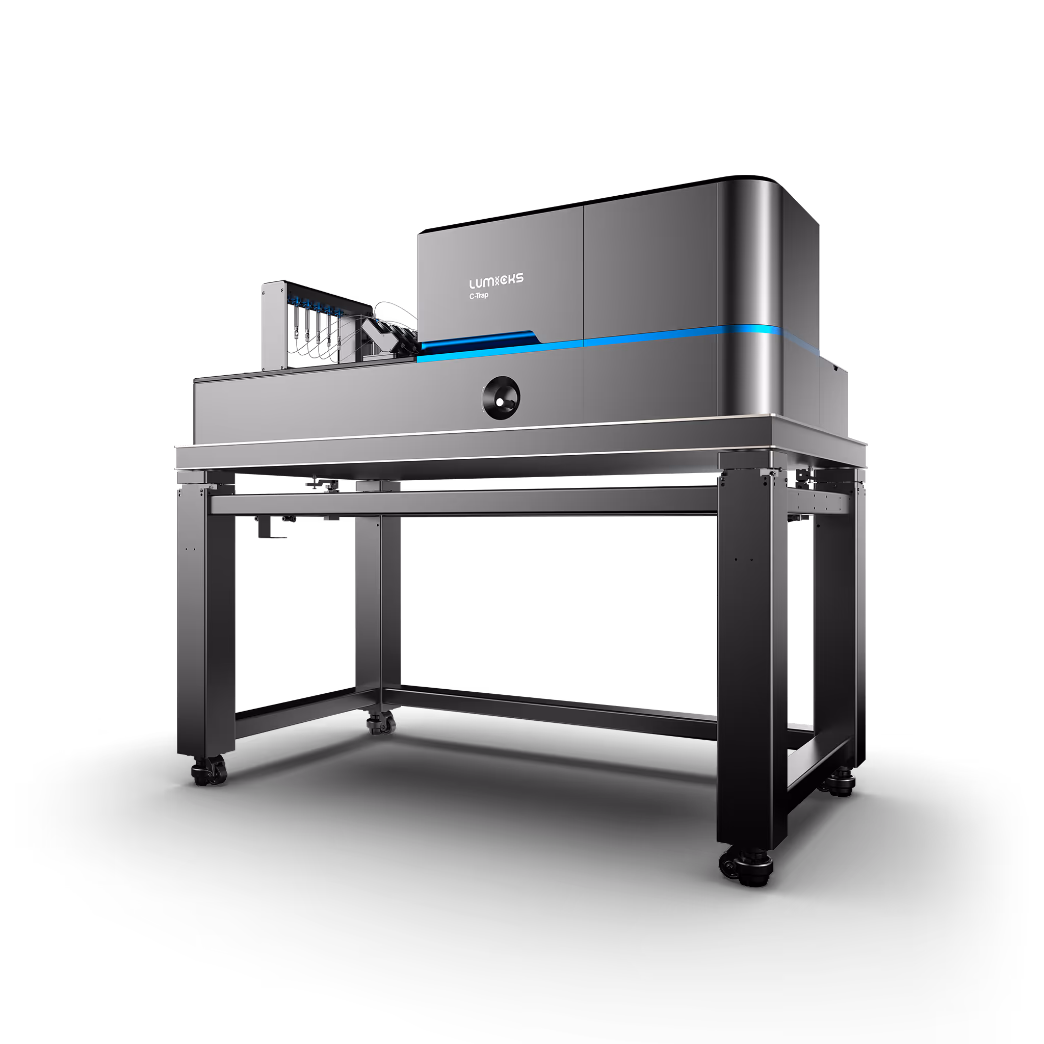
C-Trap
Molecular biology as never seen before
The C-Trap® provides the world’s first dynamic single-molecule microscope to allow simultaneous manipulation and visualization of single-molecule interactions in real time.
Technical note:
Filter CA and show 4 Latest
This shows the most recent card of each resource type filtered on Business Unit CA.
Webinar, Scientific update, Whitepaper, Application note, Brochure.
We only show 4 and we have 6 types so the 2 older ones are hidden.
In design only 1 is shown, but the rest will be loaded when published.
Button Text
Avidity and functional characterization of CD19/BCMA dual-targeting CAR-T cell designs in NHL
Webinar
July 1, 2026
01-01-20
CD19-directed CAR-T therapy has achieved remarkable clinical outcomes in patients with relapsed or refractory non-Hodgkin lymphoma (NHL). Nevertheless, a significant proportion of patients develop primary resistance or relapse, often associated with CD19 antigen loss or downregulation. Given the co-expression of CD19 and BCMA in NHL, we hypothesized that dual antigen targeting could improve therapeutic durability and mitigate antigen escape. To address this, we developed a panel of CD19/BCMA dual-targeting CAR-T cells building on our academic platforms targeting CD19 (varnimcabtagene autoleucel, ARI0001) and BCMA (cesnicabtagene autoleucel, ARI0002h). Multiple strategies were explored, including pooled mono-targeted products, co-transduction with two lentiviral vectors, bicistronic constructs, and tandem/loop CAR designs incorporating dual binding domains within a single receptor. Functional activity and avidity were assessed across varying antigen expression conditions. We found that dual CAR-T cells generated through co-transduction consistently showed superior performance relative to single-target CD19 CAR-T cells and other dual-targeting formats, particularly in models with low CD19 expression. A first-in-human phase I clinical trial (CARTDBG-01; NCT06097455) is currently underway to evaluate the safety and efficacy of ARI0003 in NHL.
Related publication: Bachiller, M., Barceló-Genestar, N., Rodriguez-Garcia, A., Alserawan, L., Dobaño-López, C., Giménez-Alejandre, M., ... & Guedan, S. (2025). ARI0003: Co-transduced CD19/BCMA dual-targeting CAR-T cells for the treatment of non-Hodgkin lymphoma. Molecular Therapy, 33(1), 317-335. https://doi.org/10.1016/j.ymthe.2024.11.028
This is some text inside of a div block.
Text Link

Enhancing efficacy against clear cell renal cell carcinoma through format-tuning of bispecific T cell engagers
Scientific update
January 29, 2025
01-01-20
This is some text inside of a div block.


Cell Avidity: a key to accelerate IND filing in cell therapy drug development
Whitepaper
July 1, 2023
01-01-20
This is some text inside of a div block.
Accelerate your cell engager discovery with high throughput measurements of Cell Avidity
Application note
June 1, 2023
01-01-20
T cells play a pivotal role in tumor immunosurveillance. Multispecific cell engagers (CEs) have been adopted in the field of immuno-oncology to redirect T cells toward cancer cells, thereby unleashing the anti-tumor potential of the patient’s immune system. CE-mediated cell binding induces T cell activation and the formation of an immunological synapse, which is a prerequisite for effective tumor cell lysis.
The strength of the initial binding events between a T cell and a tumor cell dictates the efficiency of the anti-tumor response. Assessing cell avidity, i.e. the total intercellular interaction strength between two cells, gives crucial insights into the efficacy of CEs as anti-tumor therapeutic agents.
Here, we deploy LUMICKS’ high throughput avidity measurement (HTAM) technology to measure CE-induced cell avidity in a high throughput manner. We demonstrate the assay performance characteristics, i.e. specificity, precision, and range, via CE titration experiments in the context of a Jurkat T cell model system. We find that the HTAM CA assay is suitable for candidate screening in high throughput, with high sensitivity and precision.
This is some text inside of a div block.

Cell Therapy Case Study Collection
Brochure
September 8, 2025
01-01-20
This is some text inside of a div block.
Let’s get in touch
Our team is standing by to help you