See more.
Know more.
We are LUMICKS, a life science company aiming to transform the world of immuno-oncology and molecular biology.
Cell Avidity
Revolutionize binding for the future of cell & antibody therapeutics
Imagine measuring the combined strength of cell-cell / cell-protein binding, generating direct, physiologically relevant measurements of binding in its full, dynamic complexity. To support progress in immunotherapy, we need to measure binding the way it happens—in real life, in real cells. This is Cell Avidity.
Dynamic Single-Molecule
Revealing biomolecular insights never before available
What if you could measure molecular properties and interactions while simultaneously revealing the processes in real-time? A technology that unravels the crucial and dynamic interactions taking place at the molecular level and gives you direct proof of the mechanisms involved. Our Dynamic Single-Molecule technology provides you with all of that, enabling the understanding of the root of disease development at the molecular level and accelerate therapeutic breakthroughs.

Our mission
We empower the academic and pharmaceutical communities with cutting-edge technologies to deeply understand the mechanisms of life and disease, driving the discovery and development of life-saving therapies.
What our users say
Why leading scientists trust LUMICKS technology
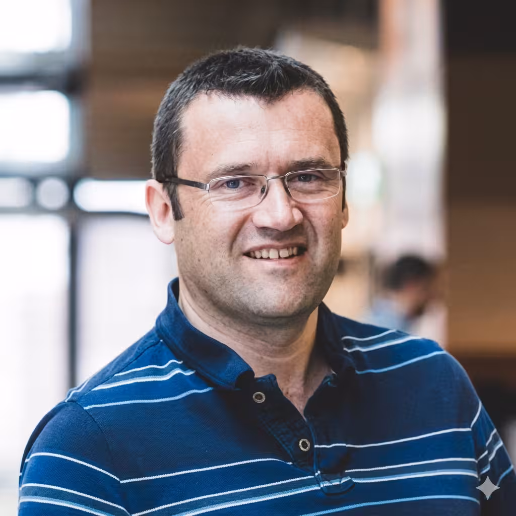
Simon studies how failures in DNA repair lead to cancer and other diseases
"I think applications like the C-Trap will become more mainstream in terms of understanding DNA transaction-based reactions."
Simon Boulton, PhD
Principal Group Leader
The Francis Crick Institute

Yufei develops CAR T cell therapy for advanced clear cell renal cell carcinoma
"Fine-tuning the cell avidity of anti-CAIX CAR T cells [enabled us to] mitigate on-target, off-tumor toxicity, [...] ensuring only CAIX-high tumor cells are killed."
Yufei Wang, PhD
Instructor of Medicine
Dana Farber Cancer Institute

Marco's team contributes to smarter, more effective treatments
"[Cell] Avidity measurement[s] will be an essential tool to screen the constructs and the products that you want to bring to the clinic."
Marco Ruella, MD
Assistant Professor of Medicine and Scientific Director of the Lymphoma Program
University of Pennsylvania
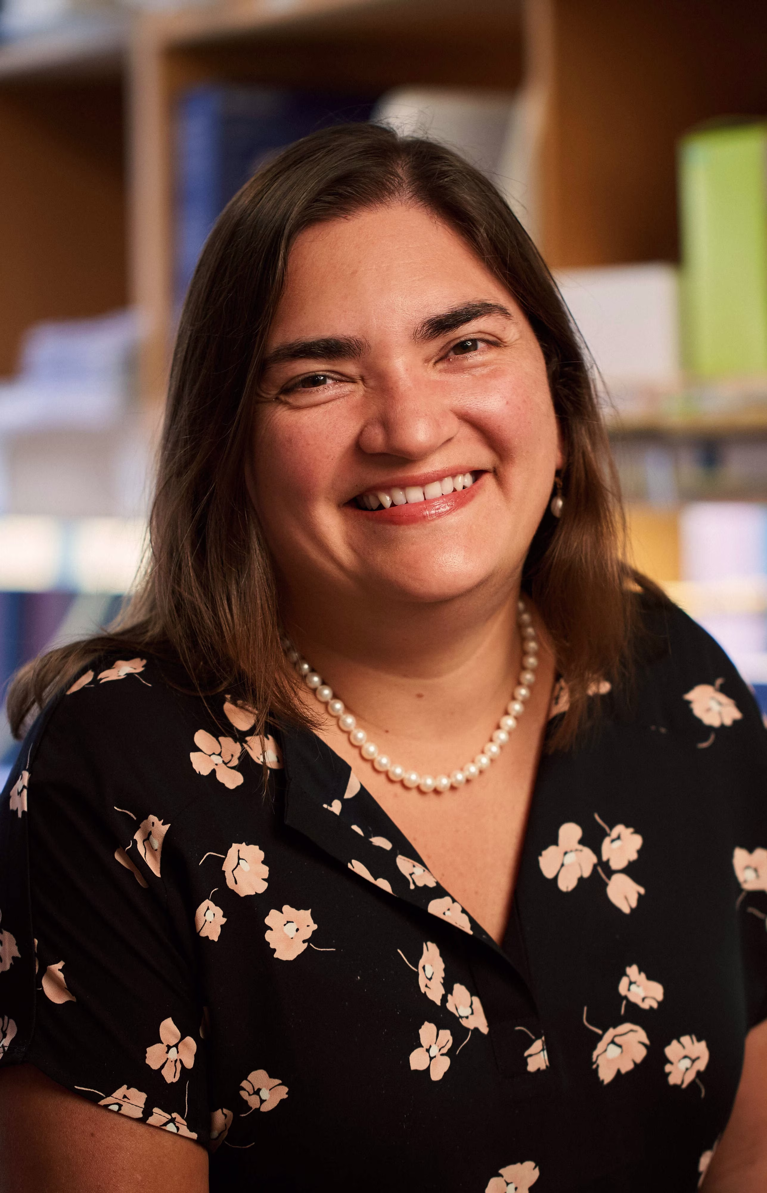
Marcela develops CAR T cell therapies to treat cancer
"Interestingly, of the pre-clinical assays we used to compare CAR constructs, only [Cell] Avidity correlated with in vivo results."
Marcela Maus, MD, PhD
Director of Cellular Immunotherapy
Massachusetts General Hospital
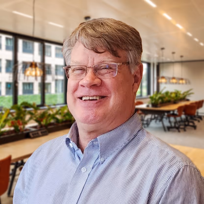
Ben's lab studies how cells repair DNA damage to prevent diseases like cancer
"The C-Trap has allowed us to ask those questions we couldn't probe before. We go down to the C-Trap room and it works first-time."
Ben Van Houten, PhD
Professor of Molecular Oncology
University of Pittsburgh
Button Text
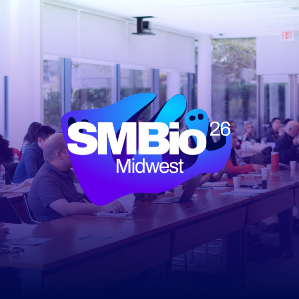
SMBio 2026 Midwest
Symposium
November 10, 2026
01-01-20
This is some text inside of a div block.


The integrated stress response promotes immune evasion through lipocalin 2
Webinar
November 2, 2026
01-01-20
This is some text inside of a div block.

The CAR Immune Synapse Interactome: How Membrane Organization Shapes CAR T–Tumor Cell Interactions
Webinar
September 14, 2026
01-01-20
This is some text inside of a div block.
.jpeg)
.png)
解析有丝分裂染色体的力学性质
Webinar
August 5, 2026
01-01-20
This is some text inside of a div block.
.png)
No items found.